01 / Alterations
Three classes of DNA alteration
Sequence variants, copy number alterations and fusions or rearrangements detectable in DNA, within the regions captured by the panel.
560 genes analyzed.
Precision oncology
A test of the tumor's DNA that can help the doctor choose the treatment. It analyzes 560 genes in a tumor sample already removed in the biopsy or surgery.


For patients and families
A test of the tumor's DNA can help your doctor choose the most suitable treatment.

DNA is the instruction manual of our cells. Each gene is a chapter. The test reads 560 genes and looks for changes that can make the tumor grow.
Somatic means “from the tumor”: the test looks for changes that arose in it. It is a test for people who have already been diagnosed with cancer, not a prevention test.
Targeted therapyMedicines that act on a specific change in the tumor. The test can guide the choice.
ImmunotherapyIt activates the body's defenses. The test can help assess whether it is an option.
Clinical trialsResearch studies with new treatments. With the test, your doctor can look for one of them.
Knowing the tumorSome changes help the doctor understand the type and the behavior of the tumor.
Is it suitable for my type of cancer? It depends on the type of tumor and the stage of treatment. Your doctor is the one who assesses this.

Not always.
Not every change has a medicine. If none shows up, that does not rule out other treatments your doctor may recommend. Your doctor reviews the result and decides with you.
The options depend on the tumor type and on the medicines approved in Brazil for it.

A report with the changes found in the tumor and what each one may mean for treatment. Take it to your appointment: your doctor explains the result and decides the next steps with you.
See a sample reportYour doctor orders the test. Without the order, the test is not performed.
The test uses the material already removed in the biopsy or surgery. It is kept by the laboratory that analyzed the biopsy. In general, another biopsy is not needed.
We explain to you or your doctor how to request this material and send it, and check beforehand whether it is suitable for the test.
Up to 15 calendar days after the sample reaches our laboratory and is accepted.
Calendar days include Saturdays, Sundays and public holidays. The time needed to gather and send the material comes before that and is not included in this count.
Talk to our team on WhatsApp or at 0800 080 0420. They will tell you about the documents, the costs and the payment options.
Health plan coverage depends on your contract, your doctor's indication and the plan's authorization.
See the health plan coverage criteria
This test analyzes only the tumor. It does not assess whether the cancer may run in the family. For that, your doctor may recommend another test, such as Neo360.
What to do now
“NeoPainel Somático or Neo360: is either one right for me?”
They will guide you on the documents, the material and the payment.
The report informs. Your doctor decides with you.
Questions about the test or the sample? Talk to our team.
Clinical indication
When the physician needs to investigate molecular alterations in the tumor that may inform treatment selection, diagnostic interpretation or assessment of clinical trials. The indication depends on the tumor type and clinical question.
Discuss the indicationClinical scope
Alteration classes that often require separate tests are assessed in the same assay, from DNA extracted from tumor tissue.
01 / Alterations
Sequence variants, copy number alterations and fusions or rearrangements detectable in DNA, within the regions captured by the panel.
560 genes analyzed.
02 / Biomarkers
Tumor mutational burden is calculated over the coding region with adequate coverage in the sample itself. Microsatellite instability is assessed at dedicated panel loci.
03 / Interpretation
Alterations are classified according to AMP/ASCO/CAP guidelines to support decisions on targeted therapies, immunotherapy and clinical trials.
Clinical use
The result should be interpreted by the treating physician together with clinical and histopathological data. Therapy selection depends on tumor type, current regulatory approvals and clinical judgment.interpreted by the treating physician

Identification of alterations that may guide the choice of targeted therapies, according to tumor type.
TMB and MSI are among the biomarkers considered when evaluating immunotherapy.
Molecular alterations that may be targeted by treatments being tested in clinical trials.
Alterations with diagnostic or prognostic significance, depending on the tumor context.
Explore the report
Understand the questions each part of the result helps answer.
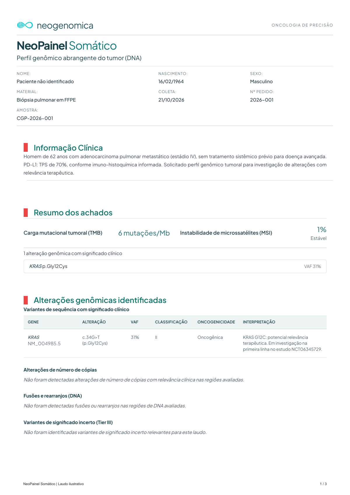
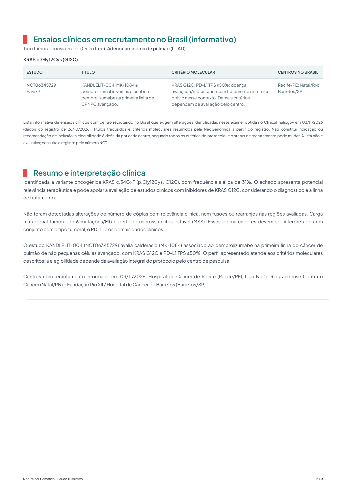
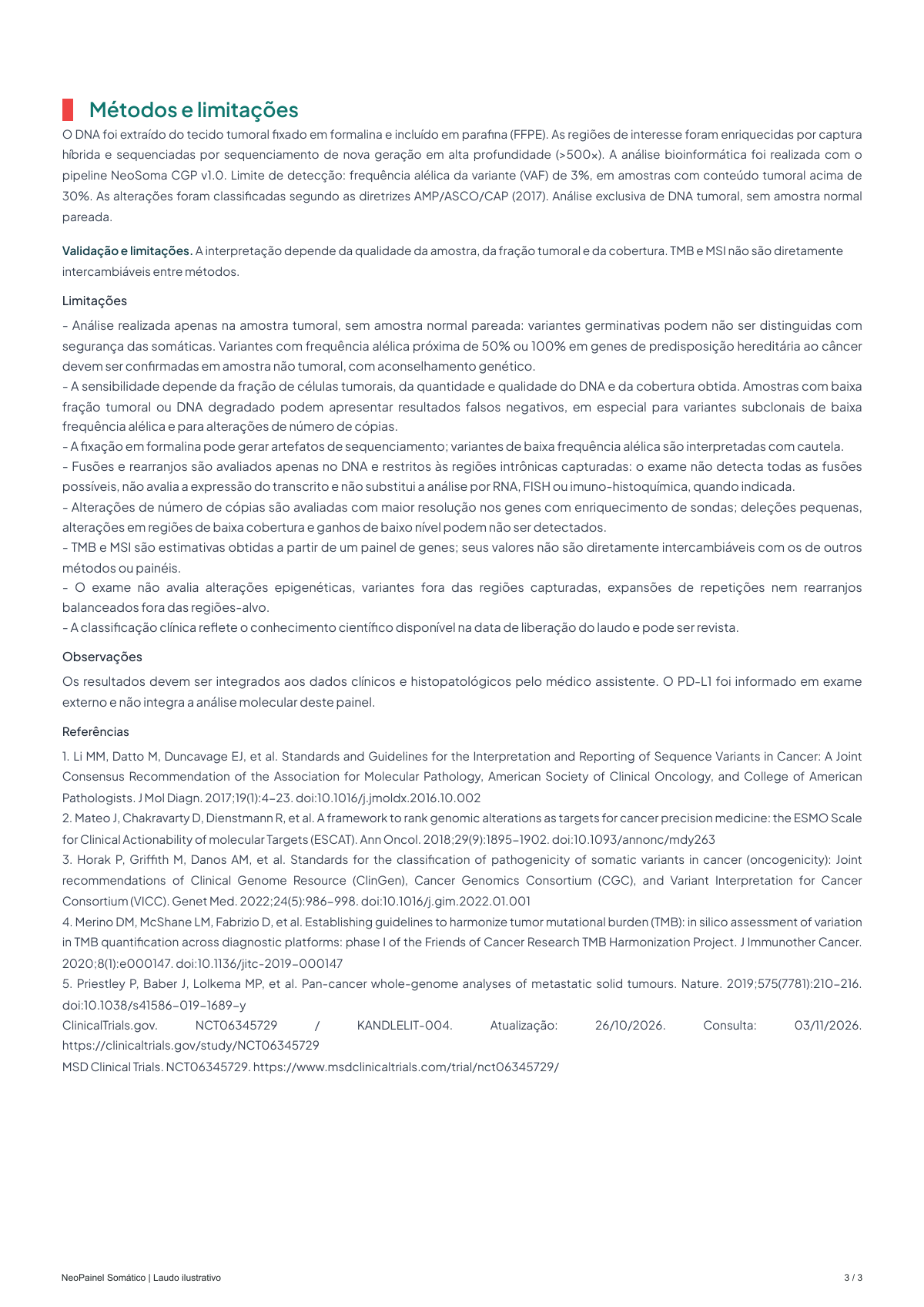
Illustrative report, not for diagnostic use.
The report format may vary according to the findings and modules assessed.
01 / Tumor profile
Not every finding has an associated therapy. The physician assesses the evidence, diagnosis and options available for that patient.
Under the AMP/ASCO/CAP guidelines, by the evidence of therapeutic, diagnostic or prognostic significance: Tier I, strong clinical significance; Tier II, potential clinical significance; Tier III, unknown significance.
The absence of a reported target does not rule out other care options. The result must be interpreted within the scope and limitations of the test.
Scope and limits
The panel covers the coding regions of 560 cancer-associated genes, with additional content for fusions, copy number and microsatellites.
High-depth sequencing (above 500x), with a detection limit of 3% VAF in samples with tumor content above 30%.
Important limitations
Panel content
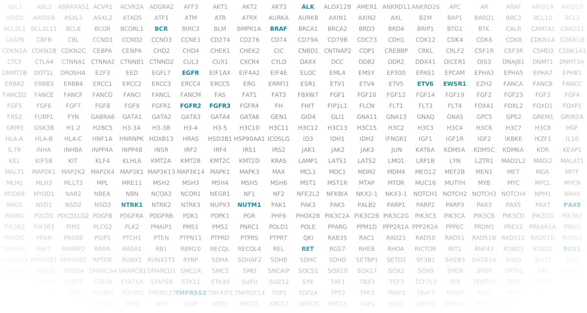
HGNC-approved gene symbols in alphabetical order, according to the current technical content of the panel.
Choice of test
Both use the 560-gene tumor panel. Neo360 adds paired tumor–normal comparison, hereditary predisposition and pharmacogenomics. The choice depends on the clinical questions to investigate.
| Feature | NeoPainel Somático |
 Neo360Learn about Neo360 |
|---|---|---|
| Tumor–normal comparison | Tumor sample only | Paired analysis from the same patient |
| Samples | Tumor: paraffin block (FFPE) or extracted DNA | Tumor and germline sample: blood, buccal swab or DNA |
| Somatic tumor profile | 560 genes, TMB and MSI | 560 genes, TMB and MSI |
| Hereditary cancer predisposition | Not included | 106 genes, from whole-genome sequencing |
| Pharmacogenomics | Not included | 6 genes and 12 drugs |
| Report | Somatic tumor profile | Integrated report in three parts |
NeoPainel SomáticoWhy germline analysis matters: an alteration found in the tumor may also be present in the normal sample; that is, it may be germline. In Neo360, comparing the two samples helps distinguish germline variants from alterations acquired by the tumor. Neo360 also assesses 106 hereditary predisposition genes, an assessment that may identify a hereditary cancer risk, with possible implications for relatives. Germline findings should be discussed in the context of clinical care and genetic counseling.
How to order
Our team provides guidance on documentation and shipping before analysis.
Medical order, tumor type, and clinical and histopathological information.
Ask the team about quantity, preservation, identification and acceptance criteria for the available material.
View technical requirementsTurnaround starts after sample acceptance, according to the test conditions.
Frequently asked questions
Documentation and references
Review the test method, scope and limitations. Interpretation references do not replace the specific technical documentation of the assay.
Frequently asked questions
A paraffin block (FFPE) with tumor tissue or DNA extracted from tumor tissue. Our team provides guidance on shipping the sample.
That is not its purpose. The test analyzes only the tumor sample, without a matched normal sample, and germline variants may not be reliably distinguished. Suggestive findings in predisposition genes should be confirmed in a non-tumor sample, with genetic counseling. Neo360 includes germline analysis.
TMB estimates the number of somatic mutations per megabase. MSI assesses microsatellite instability. Results must be interpreted with the assay limitations and clinical context; they are not directly interchangeable between methods.
No. Fusions and rearrangements are assessed in DNA, in the captured intronic regions of 14 genes. The test does not assess transcript expression and does not replace RNA, FISH or immunohistochemistry analysis when indicated.
15 calendar days, after acceptance of the sample.
Yes. The tumor type and the clinical and histopathological data guide the analysis and the interpretation of the alterations found.
Test information
Comprehensive genomic profiling of the tumor: somatic alterations in 560 cancer-associated genes, plus TMB and MSI, from tumor tissue DNA.
560 genes analyzed.
ABL1, ABL2, ABRAXAS1, ACVR1, ACVR2A, ADGRA2, AFF3, AKT1, AKT2, AKT3, ALK, ALOX12B, AMER1, ANKRD11, ANKRD26, APC, AR, ARAF, ARID1A, ARID1B, ARID2, ARID5B, ASXL1, ASXL2, ATAD5, ATF1, ATM, ATR, ATRX, AURKA, AURKB, AXIN1, AXIN2, AXL, B2M, BAP1, BARD1, BBC3, BCL10, BCL2, BCL2L1, BCL2L11, BCL6, BCOR, BCORL1, BCR, BIRC3, BLM, BMPR1A, BRAF, BRCA1, BRCA2, BRD3, BRD4, BRIP1, BTG1, BTK, CALR, CAMTA1, CARD11, CASP8, CBFB, CBL, CCND1, CCND2, CCND3, CCNE1, CD274, CD276, CD74, CD79A, CD79B, CDC73, CDH1, CDK12, CDK4, CDK6, CDK8, CDKN1A, CDKN1B, CDKN2A, CDKN2B, CDKN2C, CEBPA, CENPA, CHD2, CHD4, CHEK1, CHEK2, CIC, CNBD1, CNTNAP2, COP1, CREBBP, CRKL, CRLF2, CSF1R, CSF3R, CSMD3, CSNK1A1, CTCF, CTLA4, CTNNA1, CTNNA2, CTNNB1, CTNND2, CUL3, CUX1, CXCR4, CYLD, DAXX, DCC, DDB2, DDR2, DDX41, DICER1, DIS3, DNAJB1, DNMT1, DNMT3A, DNMT3B, DOT1L, DROSHA, E2F3, EED, EGFL7, EGFR, EIF1AX, EIF4A2, EIF4E, ELOC, EML4, EMSY, EP300, EPAS1, EPCAM, EPHA3, EPHA5, EPHA7, EPHB1, ERBB2, ERBB3, ERBB4, ERCC1, ERCC2, ERCC3, ERCC4, ERCC5, ERG, ERRFI1, ESR1, ETV1, ETV4, ETV5, ETV6, EWSR1, EZH2, FANCA, FANCB, FANCC, FANCD2, FANCE, FANCF, FANCG, FANCI, FANCL, FANCM, FAS, FAT1, FAT3, FBXW7, FGF1, FGF10, FGF12, FGF14, FGF19, FGF2, FGF23, FGF3, FGF4, FGF5, FGF6, FGF7, FGF8, FGF9, FGFR1, FGFR2, FGFR3, FGFR4, FH, FHIT, FIP1L1, FLCN, FLT1, FLT3, FLT4, FOXA1, FOXL2, FOXO1, FOXP1, FRS2, FUBP1, FYN, GABRA6, GATA1, GATA2, GATA3, GATA4, GATA6, GEN1, GID4, GLI1, GNA11, GNA13, GNAQ, GNAS, GPC5, GPS2, GREM1, GRIN2A, GRM3, GSK3B, H1-2, H2BC5, H3-3A, H3-3B, H3-4, H3-5, H3C10, H3C11, H3C12, H3C13, H3C15, H3C2, H3C3, H3C4, H3C6, H3C7, H3C8, HGF, HLA-A, HLA-B, HLA-C, HNF1A, HNRNPK, HOXB13, HRAS, HSD3B1, HSP90AA1, ICOSLG, ID3, IDH1, IDH2, IFNGR1, IGF1, IGF1R, IGF2, IKBKE, IKZF1, IL10, IL7R, INHA, INHBA, INPP4A, INPP4B, INSR, IRF2, IRF4, IRS1, IRS2, JAK1, JAK2, JAK3, JUN, KAT6A, KDM5A, KDM5C, KDM6A, KDR, KEAP1, KEL, KIF5B, KIT, KLF4, KLHL6, KMT2A, KMT2B, KMT2C, KMT2D, KRAS, LAMP1, LATS1, LATS2, LMO1, LRP1B, LYN, LZTR1, MAD2L2, MAGI2, MALAT1, MALT1, MAP2K1, MAP2K2, MAP2K4, MAP3K1, MAP3K13, MAP3K14, MAPK1, MAPK3, MAX, MCL1, MDC1, MDM2, MDM4, MED12, MEF2B, MEN1, MET, MGA, MITF, MLH1, MLH3, MLLT3, MPL, MRE11, MSH2, MSH3, MSH4, MSH5, MSH6, MST1, MST1R, MTAP, MTOR, MUC16, MUTYH, MYB, MYC, MYCL, MYCN, MYD88, MYOD1, NAB2, NBEA, NBN, NCOA3, NCOR1, NEGR1, NF1, NF2, NFE2L2, NFKBIA, NKX2-1, NKX3-1, NOTCH1, NOTCH2, NOTCH3, NOTCH4, NPM1, NRAS, NRG1, NSD1, NSD2, NSD3, NTRK1, NTRK2, NTRK3, NUP93, NUTM1, PAK1, PAK3, PAK5, PALB2, PARP1, PARP2, PARP3, PAX3, PAX5, PAX7, PAX8, PBRM1, PDCD1, PDCD1LG2, PDGFB, PDGFRA, PDGFRB, PDK1, PDPK1, PGR, PHF6, PHOX2B, PIK3C2A, PIK3C2B, PIK3C2G, PIK3C3, PIK3CA, PIK3CB, PIK3CD, PIK3CG, PIK3R1, PIK3R2, PIK3R3, PIM1, PLCG2, PLK2, PMAIP1, PMS1, PMS2, PNRC1, POLD1, POLE, PPARG, PPM1D, PPP2R1A, PPP2R2A, PPP6C, PRDM1, PREX2, PRKAR1A, PRKCI, PRKDC, PRKN, PRSS8, PSIP1, PTCH1, PTEN, PTPN11, PTPRD, PTPRS, PTPRT, QKI, RAB35, RAC1, RAD21, RAD50, RAD51, RAD51B, RAD51C, RAD51D, RAD52, RAD54L, RAF1, RANBP2, RARA, RASA1, RB1, RBM10, RECQL, RECQL4, REL, RET, RGS7, RHEB, RHOA, RICTOR, RIT1, RNF43, ROBO1, ROBO2, ROS1, RPS6KA4, RPS6KB1, RPS6KB2, RPTOR, RUNX1, RUNX1T1, RYBP, SDHA, SDHAF2, SDHB, SDHC, SDHD, SETBP1, SETD2, SF3B1, SH2B3, SH2D1A, SHQ1, SLIT2, SLX4, SMAD2, SMAD3, SMAD4, SMARCA4, SMARCB1, SMARCD1, SMC1A, SMC3, SMO, SNCAIP, SOCS1, SOX10, SOX17, SOX2, SOX9, SPEN, SPOP, SPTA1, SRC, SRSF2, STAG1, STAG2, STAT3, STAT4, STAT5A, STAT5B, STK11, STK40, SUFU, SUZ12, SYK, TAF1, TBX3, TCF3, TCF7L2, TEK, TENT5C, TERC, TERT, TET1, TET2, TFE3, TFRC, TGFBR1, TGFBR2, TMEM127, TMPRSS2, TNFAIP3, TNFRSF14, TOP1, TOP2A, TP53, TP63, TRAF2, TRAF7, TRRAP, TSC1, TSC2, TSHR, U2AF1, VAV1, VEGFA, VHL, VTCN1, WRN, WT1, XIAP, XPO1, XRCC1, XRCC2, XRCC3, XRCC5, YAP1, YES1, ZBTB2, ZBTB7A, ZFHX3, ZNF217, ZNF703, ZRSR2
Analysis of the tumor sample only, without a matched normal sample: germline variants may not be reliably distinguished from somatic ones. Variants with an allele frequency near 50% or 100% in hereditary cancer predisposition genes should be confirmed in a non-tumor sample, with genetic counseling.
Sensitivity depends on the tumor cell fraction, DNA quantity and quality, and the coverage obtained. Samples with low tumor fraction or degraded DNA may yield false-negative results, especially for low-allele-frequency subclonal variants and for copy number alterations.
Formalin fixation may cause sequencing artifacts; low-allele-frequency variants are interpreted with caution.
Fusions and rearrangements are assessed only in DNA and in the captured intronic regions: the test does not detect every possible fusion, does not assess transcript expression and does not replace RNA, FISH or immunohistochemistry analysis when indicated.
Copy number alterations are assessed at higher resolution in genes with probe enrichment; small deletions, low-coverage regions and low-level gains may not be detected.
TMB and MSI are estimates obtained from a gene panel; their values are not directly interchangeable with those of other methods or panels.
The test does not assess epigenetic alterations, variants outside the captured regions, repeat expansions or balanced rearrangements outside the target regions.
Next step
We provide guidance on the sample, documentation and limits of NeoPainel Somático before shipment.
PatientsPatient area Test results